The Purpose of this Information
This information is being provided to you in order to prepare you to make decisions about your own health care. If you should ultimately decide that surgery is the best treatment option for you, this section will help you understand what happens during an anterior cervical discectomy and will help you prepare for your role in the healing and recovery process. Read it thoroughly and answer the questions before making your final decision about your treatment options.
The Health Care Team’s Role The duty of your health care team is to:
- evaluate your condition
- establish a diagnosis
- present the various treatment options
- offer a specific treatment recommendation
- provide you with the information you need to make a decision; and then
support you in the decision you make.
The Patient’s Responsibilities
You are the only one who can decide to have surgery. It is important that you take ownership of this decision, recognizing the limitations your particular physical condition places on the potential success of each of the treatment options.
If you choose to have surgery, your physical condition and your mental attitude will determine your body’s ability to heal. You must approach your surgery with confidence, a positive attitude, and a thorough understanding of the anticipated outcome. You should have realistic goals – and work steadily to achieve those goals.
The decision to have or not to have surgery includes weighing the risks and benefits involved. You will make the final decision, so ask questions about anything you do not understand.
Since medical care is tailored to each person’s needs and differences, not all information presented here will apply to the patient’s treatment or its outcome. Seek the advice of your physician and other members of the health care team for specific information about the patient’s medical condition.
Anatomy of the Back
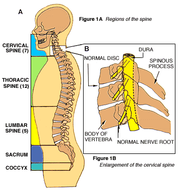
The spinal column, or backbone, consists of 33 bones (vertebrae) and can be divided into five segments (Fig. 1A). The uppermost 24 vertebrae are separated from one another by fibrous cartilage pads, called intervertebral discs (Fig. 1B), which allow the spine to bend and act as shock absorbers during activity. In the lowest part of the spine, the vertebrae are naturally fused to form the sacrum and the coccyx (tail bone).

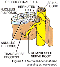
Protruding from the back of each block-like vertebral body is an arch of bone that helps to form the large, vertical spinal canal, which surrounds the spinal cord and nerve bundles (Fig. 1C, below). A fluid-filled protective membrane, the dura, covers the contents of the spinal canal from where the cord begins at the base of the brain to where it ends (in a bundle of nerve fibers known as the cauda equina).
 A pair of spinal nerves branches at each vertebral level (one to the left and one to the right), providing sensation and movement to all parts of the body. Three large bone processes arise from the vertebrae’s arch-one to each side (transverse) and one straight toward the back of the body (spinous). Strong ligaments and muscles attached to the vertebrae both support the spine and further protect the delicate spinal cord and nerves inside.
Anterior Cervical Discectomy
What is it?
Anterior cervical discectomy is an operation performed on the upper spine to relieve pressure on one or more nerve roots, or on the spinal cord. The procedure is explained by the words anterior (front), cervical (neck), and discectomy (cutting out the disc).
Why is it done?
 Neck and arm pain, among other symptoms, may occur when an intervertebral disc herniates. This happens, either suddenly with injury or slowly over time, when some of the disc’s jelly-like center (the nucleus pulposus) bulges or ruptures through its tough, fibrous outer ring (the annulus fibrosus) and presses on a nerve (Fig 1C, above).
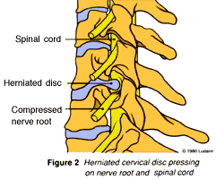
When a disc ruptures in the cervical spine, it puts pressure on one or more nerve roots (often called nerve root compression) or on the spinal cord, as seen in (Figure 2). This pressure causes symptoms in the neck, arms, and even legs. Further pressure may be caused by rough edges of bone, called bone spurs, that naturally build up around some herniated discs.
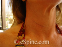
In this operation, the cervical spine is reached through a small incision in the front of your neck.
 After the soft tissues of the neck are separated, the intervertebral disc and bone spurs are removed. The space left between the vertebrae may be left open or filled with a small piece of bone. In time the vertebrae may fuse, or join together.
If used, the pre-formed bone graft may be obtained from a bone bank. It will not be rejected by your body, because it is avascular (contains no blood cells). In some circumstances, or if your surgeon prefers, the bone graft might instead be removed from your own hip through a second incision.
What happens afterwards?
Successful recovery from anterior cervical discectomy requires that you approach the operation and recovery with confidence based on a thorough understanding of each process. Your surgeon has the training and expertise to correct physical defects by performing the operation; he and the rest of the health care team will support your body’s efforts to heal its damaged tissues. Full recovery will also depend on you having a strong, positive attitude, setting small, realistic goals for improvement, and working steadily to accomplish each goal.